Hello everybody,nice to see you all again.we are learning lowerlimb in detail and we learned Lumbosacral plexus and 4 major nerves Common peroneal nerve,Tibial nerve,Femoral nerve and Obturator nerve and their clinical application.so i request you to please go through all my previous posts to gain knowledge about Anatomy.
Now we are going to learn about Femoral Triagnle and Gluteal region
FEMORAL TRIANGLE
It is present in the upper ⅓rd of the anterior aspect of the thigh.


Triangle means it has boundaries or sides.so here the boundaries of femoral triangle are -
- Base - Inguinal ligament
- Laterally- Medial border of sartorius
- Medially - Medial border of adductor longus
Even though it is called as triangle,we have to imagine the femoral triangle in 3D manner that it has floor and roof
Floor of Femoral triangle is made up of -

- Iliacus muscle
- Psoas major muscle
- Pectineus muscle
- Adductor longus muscle
Roof of Femoral triangle is made up of -

- Fascia lata is a fascia in the thigh that covers the femoral triangle
- There is opening in fascia lata that opening is called Saphenous opening this saphenous opening is covered a fascia called cribriform fascia
- the saphenous opening has a vein as content called Femoral vein and it drains venous blood from Saphenous vein and Superficial External Pudendal vessels and Superficial epigastric vessels and Superficial circumflex Iliac vessels
So these are the things that are present in roof and floor of the femoral triangle.
Now what are the contents of Femoral triangle(from lateral side to medial) -
1)Femoral nerve
2)Femoral artery
3)Femoral vein
4)Lymph nodes
Image source
These contents are arranged or present in relation to femoral sheath.so what is femoral sheath.now try to understand femoral sheath -

FEMORAL SHEATH
It is a sheath which is formed by the extension of fascia transversalis and extension of fascia iliaca which forms anterior and posterior wall respectively
The femoral nerve is not a content of femoral sheath but it is content of femoral traingle.Femoral sheath is present in femoral traingle and all the contents of femoral triangle pass through the femoral sheath except femoral nerve which passes outside of the femoral sheath.

Femoral sheath has 3 compartments -
| Lateral compartment | Intermediate compartment | Medial compartment/Femoral canal |
|---|---|---|
| Femoral artery | Femoral vein | Lymph nodes of CLOQUET/ROSENMULLER |
| Femoral branch of Genitofemoral nerve | - | - |
Lymph nodes of cloquet/rosenmuller -
In males,it drains lymph from glans penis
In females,it drains lymph from clitoris
Femoral ring
Femoral ring is base of Femoral canal

Boundaries of Femoral ring are -
- Anteriorly - Inguinal ligament
- Posteriorly - Pectineal ligament
- Medially - Lacunar ligament
- Laterally - Femoral vein
Clinical Aspects
- Femoral hernia - most commonly seen in females occurs in the femoral canal.
ADDUCTOR CANAL
It is also called Subsartorial canal because it presentbeneath sartorius muscle and Hunter's canal
You can see in the below pic

Boundaries of Adductor canal are formed by -
- Adductor magnus & Adductor longus
- Fibrous septum
- Vastus medialis
You can see boundaries of Adductor canal below image that i have drawn

The contents or structures that are passing through Adductor canal are -
- Femoral vein
- Femoral artery
- Saphenous nerve
- Nerve to vastus medialis
GLUTEAL REGION
The buttocks are called Gluteal region and we have to know what are the muscles present in this region and their nerve supply first so that we can understand the clinical aspects of it later

| Muscle | Nerve supply | Action of the muscle |
|---|---|---|
| Gluteus maximus(largest muscle) | Inferior gluteal nerve(L5,S1,S2 | Extension at hop joint |
| Gluteus medius and Gluteus minimus | Superior gluteal nerve(L4,L5,S1) | Abduction at hip joint and stabilizes the pelvis while walking |
| Gemellus superior | Nerve to obturator internus | Lateral rotation of hip joint |
| Obturator internus | Nerve to obturator internus | Lateral rotation of hip joint |
| Gemellus inferior | Nerve to quadratus femoris | Lateral rotation of hip joint |
| Quadratus femoris | Nerve to quadratus femoris | Lateral rotation of hip joint |

Clinical aspects
Gluteus medius and Gluteus minimus are the two muscles that play key role while walking.
The question is how? So lets understand this- while walking,when we lift one leg then the pelvis on that side tilt to the side of the leg that was lifted due to gravity.but it is not happening in our body why because,the Gluteus medius and Gluteus minimus muscles which are present in the opposite side will contract and pull the pelvis so that the pelvis become stabilized.so this how the these muscles help while walking.
What if these two muscles got paralysed because of injury to the Superior gluetal nerve injury?
In this case,the muscles won't contract and cannot pull pelvis and stabilize it so the pelvis get tilted to the side where leg was raised while walking or the opposite side of muscles paralysed.so the person move his upper part of his body to right side to stabilize the pelvis and to shift the center of gravity.this is called Duchenne's limp.you can understand what i said by observing below image

Trendelenburg Sign
It is clinical sign and is seen when the patient is asked to stand unassisted on each leg in turn.If pelvic drop occurs on the unsupported leg,then it is said to be positive sign. The Pelvic drop can be recognised by observing the level of the iliac crests on both sides.
We can undertsand this with the example suppose if the left gluteal muscles are weak, then the right side of the pelvis will drop when the patient stands on their left leg and we can understand trendelenberg sign by the below pic


Structures passing through Greater sciatic foramen
The two foramens Gretaer sciatic foramen and Lesser sciatic foramen are present in the pelvis and you can see them in the below pic

Above piriformis muscle -
- Superior gluteal nerve,artery and vein
Below piriformis muscle -
- Inferior gluteal nerve,artery,vein
- Nerve to obturator internus
- Nerve to quadratus femoris
- Sciatic nerve
- Posterior cutaneous nerve of thigh
- Pudendal nerve
- Internal pudendal artery and vein

Structures passing through Lesser sciatic foramen
- Pudendal nerve(S2,S3,S4)
- Internal pudendal artery and vein
- Obturator internus tendon

Iliotibial tract
It is condensed fascia lata present lateral side of thigh
Image source
Gluteus maximus and Tensor fascia lata get inserted into the Iliotibial tract
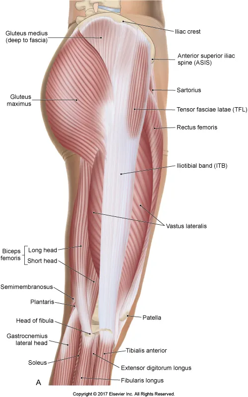
Gluteus maximus is supplied by Inferior gluteal nerve
Tensor fascia lata is supplied by Superior gluteal nerve
You can visit my previous posts of lowerlimb here -
Femoral nerve and Obturator nerve
Thanks for reading,
With regards