...Controlling Cells...Wirelessly?
Yes, you read that correctly. Scientists seek to be able to control specific cellular functions with out having to use machines and wires. It's an interesting concept that seems like science fiction. However the discovery of the protein EPG from the fish Kryptopterus bicirrhis puts humanity one step closer to making that dream a reality.

Today we will discuss some research published in the journal Nature Scientific Reports titled "Wireless control of cellular function by activation of a novel protein responsive to electromagnetic fields." In this article the authors describe their work on the EPG protein, a protein which responds to electromagnetic fields.
It has long been known that Kryptopterus bicirrhis respond to magnetic fields, with research going back as far as the 1960's describing that this magnetic sensitivity is due to a specific organ, located under the fins of the fish. [2]. This organ has a variety of calcium channels, that become activated upon exposure to any magnetic field, which results in those cells having an increased influx of calcum. It is through this calcium influx that the fish to be able to detect the magnetic field.
EPG
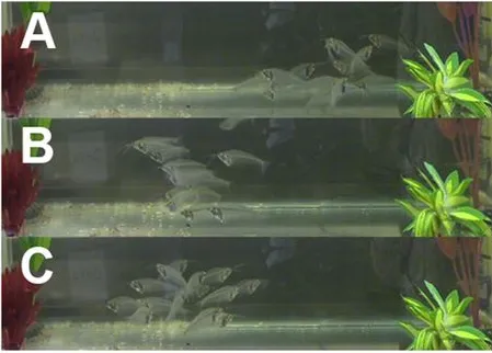
In the article we discuss today, the authors were searching for the protein which results in the activation of the calcium ion channels, and respective influx of calcium. However, they first gave a very nice demonstration of the responsiveness of the Kryptopterus bicirrhisto a magnetic field. We see in the figure to the right in A the fish are swimming in all directions happily. In B the magnetic field is turned on (on the green plant side) and the fish all swim away from it and orient themselves in the same direction. Finally in C, the magnetic field is turned off and the fish resume swimming every which way. Cool right?
To identify the protein responsible for inducing this phenomenon the researchers turned to some molecular biology trickery and extracted the mRNA from the electro responsive organ (aka all the genes being expressed) they then classified all the genes based on sequences available in the GenBank database and removed non-relevant genes. Finally they took a smaller subset and measured current response by a technique called Two-electrode voltage clamp analysis.
From this they found a small peptide of 133 amino acids that was exceedingly responsive to the current. They named this protein electromagnetic perceptive gene or EPG. They performed a variety of bioinformatic characterization steps on this peptide, however did not do the most interesting experiment that I would have done. Utilized CRISPR to knock the gene out of Kryptopterus bicirrhis and see whether or not the fish lost its perception to magnetic fields. (ah well, I guess that's work for another group to do, or another publication.)
Next Steps
Following this, the authors expressed the EPG protein in human embryonic kidney (HEK293) cells:

Here they were able to show that the EPG protein largely ends up in the membrane of the cells. The image above shows in green a stain for the protein cadherin (which is a membrane protein, that helps cells stick to one another), in blue is a DAPI stain (which stains the nucleus, as it binds to thymine bases in DNA), in red they show the fluorescently labeled EPG protein they expressed and finally they overlaid all three images together. We can see that in the EPG squares (the top is no expression of EPG and we see background fluorescence, and the bottom is when expression of the EPG is turned on) that when EPG expression is turned on, it accumulates in the same places as the cadherin, which is the cellular membrane.
The researchers then tested whether their fluorescent EPG could respond in the mammalian cells upon exposure to a magnetic field:

On the left we are looking at fluorescence prior to a magnetic field pulse, while on the right we see the fluorescence after a 10 second pulse. The darker red color is more fluorescence and as we see, fluorescence from the tagged EPG protein goes way up in the mammalian cell, after magnetic stimulation. The researchers also determined that this fluorescence coincided with a release of calcium ions from the cells (data not shown).
Can this protein be expressed in rat neurons?
It sure can, the researchers utilized lentivirus to deliver the fluorescently tagged gene to rat neurons (which we can see below on the left hand image with the green glowing neuron).

Researchers then asked whether or not the EPG protein caused a calcium response in the neurons upon exposure to a magnetic field. AKA did EPG make the neuron electromagnetic sensitive? This is what we are looking at above in the middle and right pannels. These pannels are looking at calcium concentrations through use of a calcium sensitive dye. The middle pannel is the calcium loaded neuron, and the right pannel is after 10 seconds of exposure to a magnetic field. We see that the fluorescence goes way up (it gets bright!) indicating that calcium is released from the neuron.
Yes! The expression of the protein in the neuron results in that neuron becoming sensitive to magnetic fields.
Can we make a mouse leg twitch...wirelessly?
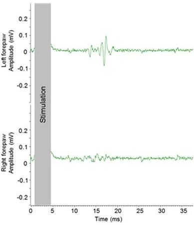
If you have learned anything from the above sections, when I ask a question in the title the answer is (at least in this piece) yes. The researchers transfected the gene into motor neurons into the right primary motor cortex of the rats (aka it should control the left limb only). They then measured the electrical potentials of both the neurons going to the right and left limbs upon exposure to a magnetic field and viola, you get what we see to the left. The left limb had an electrical response, but the right did not.
The researchers were able to wirelessly, through an electromagnetic field, control the functioning of the neurons in a specific part of the rat.
Pretty wild research, if ya ask me!